Showing posts with label microscopy. Show all posts

Monday, March 12, 2012

Measuring nucleotide concentrations inside the living cells

ResearchBlogging.org
Taking biological system apart and doing experiments in vitro is a very powerful approach. However, Nature has loads of dirty tricks up her sleeve, so doing experiments in vivo is more kosher - at least you get all the concentrations rights and will have all of the components present in the system.

Cells use a whole plethora of nucleotide-based messengers (Pesavento and Hengge, Curr. Opin. Microbiol. 2009), and following concentrations of these in vivo is something microbiologists would love to do. It is possible for some, and  c-di-GMP is an example. This nucleotide binds to numerous targets, and one of them is PilZ proteins. When binding to PilZ domain, c-di-GMP promotes a massive structural rearrangement, and this interaction can be monitored by adding a FRET pair to PilZ (Benach et al. EMBO J 2007) (Fig. 1). FRET response can be converted in c-di-GMP concentration using a calibration curve, and - viola! - c-di-GMP can be measured in the individual live cells in real time using a PilZ-GFP-based FRET detector (Christen et al. Science 2010).

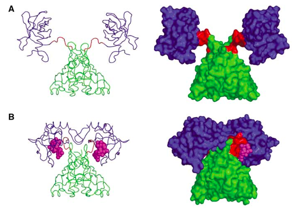

Fig. 1. PlzD: Apo (A) and in complex with c-di-GMP (figure from Benach et al. EMBO J 2007).

The problem with this approach is that is far from being universal. First, one has to have a protein that binds your target nucleotide and undergoes massive rearrangements. Second, this protein should be nice enough to work with so that you can add two GFP molecules to it to make a FRET pair, and still be able to express the protein for in vitro work (one needs to calibrate the FRET response, right?). In the case of stringent response there seem to be no such proteins for detection of my favorite nucleotide, ppGpp... too bad!

Well, there seems to be a new method out there, and this one holds great promise. In their recent Science paper Paige and colleagues use an RNA-based FRET pair using RNA mimic of GFP (Paige et al. Science 2011) combined with a small-molecule-specific aptamer (Fig. 2). When the ligand binds, RNA forms a stable structure and FRET is on! They have followed in E. coli concentrations of two molecules - ADP and SAM. However, aptamers can be evolved for other targets, and this makes this method potentially applicable for detecting whatever molecule that picks your fancy.


Fig. 2. Schematic representartion of the aptamer-based FRET sensor for in vivo detection of small molecules (figure from Paige et al. Science 2012).

References:

Paige JS, Nguyen-Duc T, Song W, & Jaffrey SR (2012). Fluorescence imaging of cellular metabolites with RNA. Science (New York, N.Y.), 335 (6073) PMID: 22403384

Paige JS, Wu KY, & Jaffrey SR (2011). RNA mimics of green fluorescent protein. Science (New York, N.Y.), 333 (6042), 642-6 PMID: 21798953 Christen M, Kulasekara HD, Christen B, Kulasekara BR, Hoffman LR, & Miller SI (2010). 


Matthias Christen, Hemantha D Kulasekara, Beat Christen, Bridget R Kulasekara, Lucas R Hoffman, and Samuel I Miller (2010) Asymmetrical distribution of the second messenger c-di-GMP upon bacterial cell division. Science (New York, N.Y.), 328 (5983), 1295-7 PMID: 20522779

 Benach J, Swaminathan SS, Tamayo R, Handelman SK, Folta-Stogniew E, Ramos JE, Forouhar F, Neely H, Seetharaman J, Camilli A, & Hunt JF (2007). The structural basis of cyclic diguanylate signal transduction by PilZ domains. The EMBO journal, 26 (24), 5153-66 PMID: 18034161

Wednesday, December 14, 2011

Single molecule tracking fluorescence microscopy in mitochondria reveals highly dynamic but confined movement of Tom40

Most of the mitochondrial proteins are imported from the cytoplasm, with only a small fraction (about 1%) encoded in the mitochondrial genome. Import is mediated by two complexes: TOM (transporter outer membrane) and TIM (transporter inner membrane). We have a pretty good idea about the players involved in mitochondrial protein import, but we have very little idea about the dynamics of TOM/TIM movement in the mitochondrial membrane.

We tried addressing this question using single molecule fluorescent microscopy in isolated yeast mitochondria. What we see is that Tom40, the central component of TOM complex, is highly confined (i.e. restricted in terms of aerea it can sample) but within its confinement it moves pretty rapidly.

References:

Kuzmenko et al., Scientific Reports (2011) 1:95

Thursday, August 4, 2011

Observing protein synthesis inside the living mammalian cell

ResearchBlogging.org


Biologists really love seeing things for themselves. Take for instance the central dogma: DNA - RNA - protein. It all well and good when represented as childish-looking blobs fooling around and passing amino acids one to each other, but how about it actually happening in the real 3D cell stuffed with other goodies? Well, obviously, people tried looking into the question.

The first option is you can to label fluorescently some components of the machinery - ribosomes, mRNAs, factors - and plonk the bug under the microscope. The problem here is that you can't really see what these components are doing - are they idle? are they active? are they so fucked up by adding the fluoresecnt label that now they are being rapidly degraded? One possible solution is to interfere with the cell inhibiting some crucial step the component of interest is involved in and see what happens to the distribution of the labeled component. For instance, one can add an antibiotic. The problem with this approach is that you a) interfere with the system b) interfere with the system c) antibiotics are often fluorescent, so you interfere with the system even more.

One solution that is often used in vitro for converting fluorescence signal into the distance signal is by using FRET, Förster resonance energy transfer (for an excellent review of FRET-based single molecule investigations of translation in vitro see Blanchard). The idea behind FRET is that one uses two fluorofores at the same time. Fluorofore 1 is excited at wavelength λ1, and emits at λ2. Fluorofore 2 is adsorbs at λ2 and emits at λ3. In order for all this cascade to work, the two fluorofores have to be very, very close because efficiency of FRET decreases with distance with an inverse 6th power law.

Now, back to observing translation in vivo. Barhoom at al. exploited the FRET strategy in their piece that just came out in NAR. They used a FRET pair consisting of two labeled tRNAs. These two are getting very close on the ribosome, generating a FRET signal (a strategy recently used in vitro by Uemura at al.). And they are getting close on the ribosome only if they are actively engaged in translation (Fig. 1). Therefore by looking at the tRNA FRET Barhoom and colleagues can observe spots of active translation inside the cell under different conditions, such as viral infection etc.

The paper is open access, so do take advantage of that.


Fig. 1. Two labeled tRNAs are constituting a FRET pair. When they are in close proximity on the ribosome they produce a FRET signal. When floating about in the cytoplasm, they are too far to generate FRET.


PS: this post is part of the MolBio carnival Nr 14!

References:

Blanchard SC (2009). Single-molecule observations of ribosome function. Current opinion in structural biology, 19 (1), 103-9 PMID: 19223173

Barhoom S, Kaur J, Cooperman BS, Smorodinsky NI, Smilansky Z, Ehrlich M, & Elroy-Stein O (2011). Quantitative single cell monitoring of protein synthesis at subcellular resolution using fluorescently labeled tRNA. Nucleic acids research PMID: 21795382

Uemura S, Aitken CE, Korlach J, Flusberg BA, Turner SW, & Puglisi JD (2010). Real-time tRNA transit on single translating ribosomes at codon resolution. Nature, 464 (7291), 1012-7 PMID: 20393556

Friday, May 27, 2011

Photoactivatable organic fluorophores for in vivo imaging

Imaging techniques heavily rely on the fluorophores they use. GFP was a great breakthrough because it allowed labeling specific targets, photoconvertable GFP variants were a great breakthrough because they allowed generation of single fluorescent molecules and this lead to development of a whole array of tracking and superresolution techniques.

However, GFP variants were always pretty bad fluorophores as compared to organic dyes due to their low quantum yield, pH and redox-potential sensitivity. Organic dyes, good as they are, had issues with selective labeling and thus were mostly used in vitro.

It seems that it might be about to change.  Lee at al. report a novel strategy for labeling proteins of interest in vivo with organic dyes. They use HaloTag technology by Promega and label a whole bunch of proteins both in mammalian cells and bacteria with photoactivatable azido DCDHF labels.

And it works beautifully. The only problem is that HaloTag is even bigger than GFP - 33 kD. That's a lot.

References:

Lee at al. Superresolution imaging of targeted proteins in fixed and living cells using photoactivatable organic fluorophores. JACS 2010 vol. 132 (43) pp. 15099-15101 PIMD 20936809

Lord at al. Azido push-pull fluorophores photoactivate to produce bright fluorescent labels. J Phys Chem B 2010 vol. 114 (45) pp. 14154-14167 PIMD 19860443

Monday, May 9, 2011

Antibiotics affecting ribosome's protein composition

This post was chosen as an Editor's Selection for ResearchBlogging.org


Antibiotics kill bugs; and about a half of them are doing so by messing up translation. That usually means that the ribosome is stalled at a certain step, be it initiation or elongation or ribosomal recycling.

But it is not always just that. Sometimes antibiotics also mess up the ribosome itself  and affect its composition.

Exhibit A: kasugamycin, an antibiotic that inhibits translation initiation in bacteria by interfering with binding of the the initiator tRNA. Amazingly enough, treatment with kasugamycin results in dramatic change in the ribosomal composition which is in turn changing ribosome's functional properties. Several proteins dissociate from the small ribosomal subunit (S1, S2, S6, S12, S18 and S21) which turns the 70S ribosome into a 61S kasugamycin particle. Ribosomal protein S1 is of particular interest here, because it is very important for the mRNA:ribosome interactions and is responsible for  A/U rich sequences acting as translational activators.

The S61 particle loses the ability to translate mRNAs with Shine-Dalgarno sequences, while being able to translate leaderless mRNAs, that is the ones starting directly with the initiation codon at the 5'. These leaderless mRNAs can be translated without the help of any initiation factors, by bacterial and eukaryotic ribosomes alike, so it is no surprise that S61 particles, even though compromised can still translate these messages.

What is particularly interesting in the kasugamycin story, it is that loss of the ribosomal proteins can be reconstituted in vitro by simply mixing the drug with the 70S. This means that the effect is direct rather than mediated by the assembly process (see below for an example of the latter effect).

Exhibit B: chloramphenicol and erythromycin. These antibiotics cause defects of the ribosomal assembly, and they seem to be doing so by interfering with the expresion levels of different ribosomal proteins. Here we have Liebig's barrel in action: you interfere with levels of many components you need to have and end up running out of one, the limiting one.

All of the above is highly relevant for people using antibiotics as tools, for instance in microscopy. Chloramphenicol and kasugamycin are widely used to inhibit translation (for instance here and here). It's worth remembering that they are doing much more than that while interpreting your results. Sometimes the tool you use can have much more complicated character than one would anticipate, as I discussed here.

References:

Wilson DN (2009). The A-Z of bacterial translation inhibitors. Critical reviews in biochemistry and molecular biology, 44 (6), 393-433 PMID: 19929179

Schluenzen F, Takemoto C, Wilson DN, Kaminishi T, Harms JM, Hanawa-Suetsugu K, Szaflarski W, Kawazoe M, Shirouzu M, Nierhaus KH, Yokoyama S, & Fucini P (2006). The antibiotic kasugamycin mimics mRNA nucleotides to destabilize tRNA binding and inhibit canonical translation initiation. Nature structural & molecular biology, 13 (10), 871-8 PMID: 16998488

Schuwirth BS, Day JM, Hau CW, Janssen GR, Dahlberg AE, Cate JH, & Vila-Sanjurjo A (2006). Structural analysis of kasugamycin inhibition of translation. Nature structural & molecular biology, 13 (10), 879-86 PMID: 16998486

Kaberdina AC, Szaflarski W, Nierhaus KH, & Moll I (2009). An unexpected type of ribosomes induced by kasugamycin: a look into ancestral times of protein synthesis? Molecular cell, 33 (2), 227-36 PMID: 19187763

Siibak T, Peil L, Dönhöfer A, Tats A, Remm M, Wilson DN, Tenson T, & Remme J (2011). Antibiotic-induced ribosomal assembly defects result from changes in the synthesis of ribosomal proteins. Molecular microbiology, 80 (1), 54-67 PMID: 21320180

Nevo-Dinur K, Nussbaum-Shochat A, Ben-Yehuda S, & Amster-Choder O (2011). Translation-independent localization of mRNA in E. coli. Science (New York, N.Y.), 331 (6020), 1081-4 PMID: 21350180

Tzareva NV, Makhno VI, & Boni IV (1994). Ribosome-messenger recognition in the absence of the Shine-Dalgarno interactions. FEBS letters, 337 (2), 189-94 PMID: 8287975

Andreev DE, Terenin IM, Dunaevsky YE, Dmitriev SE, & Shatsky IN (2006). A leaderless mRNA can bind to mammalian 80S ribosomes and direct polypeptide synthesis in the absence of translation initiation factors. Molecular and cellular biology, 26 (8), 3164-9 PMID: 16581790

Montero Llopis P, Jackson AF, Sliusarenko O, Surovtsev I, Heinritz J, Emonet T, & Jacobs-Wagner C (2010). Spatial organization of the flow of genetic information in bacteria. Nature, 466 (7302), 77-81 PMID: 20562858

Tuesday, February 1, 2011

photoconvertable GFP variants for SPT

Many different fluorescent labels are used for super-resolution imaging, and photoconvertable GTP variants are probably the most widely used. And again, there are many photoconvertable GFP variants: PATagRFP, PA-GFP, Dendra, EosFP and many others.

What makes a good photoconvertable GFP for single particle tracking (SPT)? Well, all the things that make a good label (bright, stable, fast to mature, small, inert, this convenient and nicely separated adsorbance and emission spectra - the obvious stuff) and one more thing that is not so obvious.

Photoconvertable label should not convert by itself. It should have a stable dark state and conversion should be strictly upon illumination by the photo-converting light. Otherwise creating single molecules for SPT is hard - they just pop into existence by themselves! In than sense EosFP is much better than Dendra.

Sounds obvious, but it wasn't to me...

Increasing signal-to-noise ratio by use of highly inclined thin illumination beam

Signal-to-noise ratio (SNR) is a crucial parameter in imaging. In order to improve it researchers use different tricks, optimizing the labels and reducing the background.

Here is one interesting imaging trick called Highly Inclined and Laminated Optical sheet (HILO) microscopy. Using highly inclined thin imaging beam it is possible to increase SNR dramatically.

In a sense this approach is similar to TIRF, where illuminating light is approaching the sample at a critical angle, so that only molecules very close to the coverslip surface are illuminated by the evanescent wave. The problem with TIRF is exactly that - only molecules very close to the surface can be imaged, which is fine for working with membrane proteins, but not so for the cytosolic ones.

HILO can do just that: instead of illuminating only the molecules in close proximity to the coverslide (as TIRF does) it illuminates a thin slice cutting through the cell at an angle.

References:

Tokunaga et al. Nature Methods 2008, 5 (2) pp. 159-161 PIMD 18176568

Wednesday, January 19, 2011

Single Particle Tracking (SPT) in vivo: interpretation of trajectories

Using a single particle tracking approaches it is possible to approach diffusion trajectories with millisecond time resolution. Analyzing these and figuring out what exactly is happening in the cell is a rather complex undertaking. Here are two papers discussing analysis of particle diffusion in vivo:

1. van den Wildenberg et al. Biopolimers 2011 PIMD 21240922

Use of tensors for describing asymmetry of diffusion in the cell (i.e. along the long axis we have less confinement than along the short axis, thus mean square displacement (MSD) plateau is different). Modeling of different confinement geometries (tube, cube, sector) + adding repulsive or attractive potentials describing interactions between the particle and environment.

2. Hall and Hoshino Biophys Rev 2010 PIMD 21088688

Focuses on protein diffusion in the bacterial membrane, comparing experimental data for TatA diffusion with modeling. Again, confinement effects are discussed - SPT data are 2D projection of the 3D diffusion. They model random walk on the surface of bacterial cell was in the same way as Deich at al. PNAS 2004 PIMD 15522969.

Geometric constraints and 2D-to-3D projection affect MSD and CPD (cumulative probability distributions), but not enough to explain thir experimental data.

In order to account for the experimental deviations unexplainable by geometry effects they assume existence of slow and fast particles and add an estimate of the localization precision, and now they get a nice agreement with the experimental data. Roughly 50 / 50 slow and fast.

Effect of localization precision on SPT was earlier discussed in Martin et al. Biophys J. 2002 PIMD 12324428 story: "Apparent subdiffusion inherent to single particle tracking".

Thursday, January 13, 2011

Playing chicken, single molecule

ResearchBlogging.org

Many things happen to DNA. Proteins bind, slide along, dissociate. Sometimes they bump into each other, and then... what happens then?

This was exactly the question adressed in Finkelstein at al., Nature 2010. They were particularly interested in a bacterial protein called RecBCD, which is a powerful helicase. Using single-molecule microscopy they
had a look at what happens when RecBCD rams into some other protein.

And they had several to look at. RNA polymerase was the first one, and this is a formidable roadblock, and yet - RecBCD pushed it off the DNA with ease. Here are the actual images:

RecBCD sliding RNA polymerase along the DNA (76.5 %):


RecBCD ejecting RNA polymerase (8.5 %):


And RecBCD being stalled by RNA polymerase (rare events!) (15 %):


Then they tried other proteins, EcoRI endonuclease and lac repressor, and again, RecBCD was victorious over and over. Only the mighty nucleosome (never mind that it is a eukaryotic protein and it never met RecBCD before!) managed to put up a fight - 24% of the head-on collisions resulted with RecBCD being stalled, 11% resulted in nuceosome ejection and in remaining 65% RecBCD was sliding the nucleosome along the DNA.

What next? Well, now authors are preparing for experiments on RecBCD head-on collisions with Higgs bosons in the LHC.  I bet on RecBCD.

PS: it is all very convenient that the authors happened to study RecBCD - neat story, their protein is stronger than all the others, yay! But wat if they would have started with some lame one, like PNA polumerase? But hey! I never write my results in the order I get them... do you? Did THEY?

References:

Finkelstein IJ, Visnapuu ML, & Greene EC (2010). Single-molecule imaging reveals mechanisms of protein disruption by a DNA translocase. Nature, 468 (7326), 983-7 PMID: 21107319

Monday, December 27, 2010

Double life of bacterial elongation factor EF-Tu

ResearchBlogging.org

Protein biosyntheses is performed by the ribosome and is assisted by a large number of accessory molecules, with several of them belonging to the GTPase class. EF-Tu is a GTPase which is responsible for bringing the amynoacyl-tRNA to the ribosome, and it is one of the most abundant proteins in bacteria.

So abundant, in fact, that it is impossible to explain this abundance by EF-Tu's role in translation. And indeed, recent microscopy investigations has shown how EF-Tu works as a part of bacterial cytosceleton.

EF-Tu works together with another bacterial structural protein, actin-like protein MreB, which froms spirals underneath bacterial membrane. In this tandem EF-Tu leads the formation of more dynamic MreB filaments.

In eucaryotes eEF1A does the job of EF-Tu and delivers the tRNA. It is involved in interactions with the cytosceleton just as it its bacterial counterpart, but instead of MreB it interacts with the filamentous actin, causing it to form bundles.

Bunai F, Ando K, Ueno H, & Numata O (2006). Tetrahymena eukaryotic translation elongation factor 1A (eEF1A) bundles filamentous actin through dimer formation. Journal of biochemistry, 140 (3), 393-9 PMID: 16877446

Defeu Soufo HJ, Reimold C, Linne U, Knust T, Gescher J, & Graumann PL (2010). Bacterial translation elongation factor EF-Tu interacts and colocalizes with actin-like MreB protein. Proceedings of the National Academy of Sciences of the United States of America, 107 (7), 3163-8 PMID: 20133608

Vats P, & Rothfield L (2007). Duplication and segregation of the actin (MreB) cytoskeleton during the prokaryotic cell cycle. Proceedings of the National Academy of Sciences of the United States of America, 104 (45), 17795-800 PMID: 17978175

Tuesday, December 14, 2010

GFP-based quantification of proteins in the cell - don't try that at home!

ResearchBlogging.org

This is something that come out of our tracking experiments in J. Elf lab. I guess we will never publish any quantitive account of it, so it guess it constitutes perfect blogging material.

GFP and its derivatives are widely used to label and track proteins in live cells. This way people follow localization of the protein of interest and accessing changes its concentration.

And here is where the fun begins. GFP has a fluorofor that is bringt only in a) oxidized b) deprotonized state. Therefore redox potential and pH of the cellular environment have profound effect on the brightness of GFP. Cultural media per se can change GFP behavior dramatically, and this is something outside of the cell...

Another problem with GFP is that it can radically affect stability of the fusion protein. We are not even talking about function here, we are talking about the number of molecules per se.

While struggling with our Dendra2 GFP variant and RelA_Dendra2 fusion in vivo we observed all these problems constantly. Unhappy cells were dark, with GFP in the dark state (pH? redox? you simply don't know!) and the numbers of proteins you detect were invariably much lower than what is estimated for the wt, un-tagged RelA.

But all this still does not stop brave souls from using GFP for single molecule quantification of bacterial proteins en masse. Whole library of YFP (yellow fluorescent protein) fusions was created and their copy number as well as diffusion characteristics were analyzed, and then systems biology happened to the dataset. No one knows what exactly does it all mean and how the numbers of detected YFP-fused proteins relate to numbers of the wt proteins, but this is details.

References:

Bogdanov AM, Bogdanova EA, Chudakov DM, Gorodnicheva TV, Lukyanov S, & Lukyanov KA (2009). Cell culture medium affects GFP photostability: a solution. Nature methods, 6 (12), 859-60 PMID: 19935837

Taniguchi Y, Choi PJ, Li GW, Chen H, Babu M, Hearn J, Emili A, & Xie XS (2010). Quantifying E. coli proteome and transcriptome with single-molecule sensitivity in single cells. Science (New York, N.Y.), 329 (5991), 533-8 PMID: 20671182

Brian P. English, Arash Sanamrad, Stoyan Tankov, Vasili Hauryliuk, & Johan Elf (2010). Tracking of individual freely diffusing fluorescent protein molecules in
the bacterial cytoplasm arXiv q-bio arXiv: 1003.2110v1