Wednesday, December 14, 2011

Single molecule tracking fluorescence microscopy in mitochondria reveals highly dynamic but confined movement of Tom40

Most of the mitochondrial proteins are imported from the cytoplasm, with only a small fraction (about 1%) encoded in the mitochondrial genome. Import is mediated by two complexes: TOM (transporter outer membrane) and TIM (transporter inner membrane). We have a pretty good idea about the players involved in mitochondrial protein import, but we have very little idea about the dynamics of TOM/TIM movement in the mitochondrial membrane.

We tried addressing this question using single molecule fluorescent microscopy in isolated yeast mitochondria. What we see is that Tom40, the central component of TOM complex, is highly confined (i.e. restricted in terms of aerea it can sample) but within its confinement it moves pretty rapidly.

References:

Kuzmenko et al., Scientific Reports (2011) 1:95

Friday, December 2, 2011

long memories of RelA

Enzymes have their cycles, catalytic ones: bind a substrate, catalyse some sort of reaction, release the product... then do it again. These cycles have memory effects: long turnover is likely to be followed by another long one, and short one is likely to be followed by another short one. This makes total sense: efficient act of catalysis is possible only when appropriate conformation is achieved and all the residues are aligned as they should be... and that is a recipe for one more efficient round!

Now let us look at RelA. Based on in vivo single molecule tracking investigations we recently proposed a model of RelA catalytic cycle: it sits on the ribosome, gets activated by arrival of deacylated tRNA to the A site, falls off and performs multiple acts of ppGpp synthesis from ATP and GDP. Importantly, RelA must go through the ribosome-bound stage in order to get activated. This seems to be an extreme cause of memory effects - while active off the ribosome, RelA remembers the activation event that happened on the ribosome!

It is quite a scary thought... what else does it remember? Does it remember the moment I started working on it? Well, surely not, even I don't remember that moment any more. Or may be I am just blocking out that memory.

References:

H. Peter Lu, Phys. Chem. Chem. Phys (2011), 13 pp. 6734-6749, PIMD 21409227

Brian P. English et al., PNAS (2011), 108(13) pp. E365-373, PIMD 21730169

Active role of the stringent response in antibiotic tolerance

From time to time we try killing bacteria with antibiotics. Most of the bugs die, but not all. These survivors fall into two categories: resistant bugs and tolerant bugs. Resistant bugs have specific mechanisms counteracting the drug: mutations in the target site, enzymes destroying the antibiotic, etc. Tolerant bugs are not getting killed using some more general approach, such as forming a biofilm efficiently shielding them from contact with the drug or shutting down its biosynthetic activity and waiting for the better days to come.

The stringent response is a mechanism rewiring the bacterial physiology under stress. It changes many things simultaneously, and, not surprisingly, functionality of the stringent response is linked to antibiotic tolerance. However, the big question here is the nature of this link: do bugs need functional stringent response in order to tolerate the drug just because relaxed bugs do not shut down their growth when needed and die, or does the stringent response induce production of certain specific enzymes protecting from the drug?

Recent report by Nguyen and colleagues seems to settle this question. Using series of in vivo experiments with E. coli knock-out strains deficient either in stringent response per se (knock-outs of RelA and SpoT) or in down-stream stringent response-regulated targets they show that the main source of antibiotic tolerance is not a general biosynthetic shut-down. Specifically, they identify two genes induced during the stringent response - superoxide dismutase (SOD) and catalase - to be crucial for bacterial survival in the presence of antibacterials.  What these do, they protect the bug from the hydroxyl radical. And build-up the latter was recently identified as a common mechanism causing the cell death during treatment by different unrelated antibacterials

References:

Nguyen at al., Science (2011) 334, pp. 982-986 PIMD 22096200

Kohanski et al. Cell (2007) 130, pp. 797-810 PIMD 17803904

Thursday, November 3, 2011

DksA and ppGpp regulate transcription of both rRNA and r-proteins

During the stringent response ppGpp together with a small protein DksA bind to the RNA polymerase and down-regulate transcription of the rRNA genes. It makes sense - there is no need for more ribosomes if there are not enough amino acids. However, ribosomes consist not only of rRNA but also of ribosomal proteins, and if the cell stops making rRNA it would make sense to stop making the ribosomal proteins as well.

And it turnes out that ppGpp and DksA do that too. Makes total sense, again. It is quite rare that something about the stringent response makes total sense, but there you are.

References:

Lemke, J. J., Sanchez-Vazquez, P., Burgos, H. L., Hedberg, G., Ross, W., & Gourse, R. L. (2011). Direct regulation of Escherichia coli ribosomal protein promoters by the transcription factors ppGpp and DksA. Proceedings of the National Academy of Sciences of the United States of America, 108(14), 5712–5717.

Wednesday, August 10, 2011

The RelA/SpoT Homolog (RSH) Superfamily: Distribution and Functional Evolution of ppGpp Synthetases and Hydrolases across the Tree of Life

Stringent response is run by the RSH (RelA / SpoT Homologue) proteins, but there are more RSHs then just these two. Usually researchers were finding them using an ad hoc approach: take your favorite bug you worked with for 10 years, blast its genome with RelA gene, find anything that looks like RelA, test  it.

Finally there is a proper analysis of RSHs across the tree of life: The RelA/SpoT Homolog superfamily: distribution and functional evolution of ppGpp synthetases and hydrolases across the tree of life by Atkinson GC, Tenson T, Hauryliuk V, PLoS ONE, 6(8): e23479. 

Here is the take home message:
  • there are loads of different RSHs out there: we identified 30 subgroups! 
  • all the RSHs out there are now are classified (for now, that is. New genomes are coming out every day, damn the progress!).
  • Archaea, Bacteria, Eucaryotes: they all have RSHs. I repeat: Archaea too.
  • there are the long RSHs (i.e. Rel, RelA and SpoT) and there are the short ones. 
  • The short ones have either ppGpp synthesis or ppGpp hydrolysis domain. The long ones have both, but not always both are functional.
  • by comparing the long ones vs the short ones we identified residues potentially involved in the inter-domain cross-talk in the long ones (the short ones have only one domain thus there is no cross talk there!).
The bottom line: if you work on a strange RSH protein from a strange bug, do check out our paper. 
Fig. 1.  Maximum likelihood phylogeny of the ppGpp hydrolase domain.  Subgroups are labeled and shading behind the branches shows the most common domain structure observed for those groups, as per the legend in the inset box. Symbols on branches indicate bootstrap support, as per the inset box.

Hurray to Gem

Thursday, August 4, 2011

Observing protein synthesis inside the living mammalian cell

ResearchBlogging.org


Biologists really love seeing things for themselves. Take for instance the central dogma: DNA - RNA - protein. It all well and good when represented as childish-looking blobs fooling around and passing amino acids one to each other, but how about it actually happening in the real 3D cell stuffed with other goodies? Well, obviously, people tried looking into the question.

The first option is you can to label fluorescently some components of the machinery - ribosomes, mRNAs, factors - and plonk the bug under the microscope. The problem here is that you can't really see what these components are doing - are they idle? are they active? are they so fucked up by adding the fluoresecnt label that now they are being rapidly degraded? One possible solution is to interfere with the cell inhibiting some crucial step the component of interest is involved in and see what happens to the distribution of the labeled component. For instance, one can add an antibiotic. The problem with this approach is that you a) interfere with the system b) interfere with the system c) antibiotics are often fluorescent, so you interfere with the system even more.

One solution that is often used in vitro for converting fluorescence signal into the distance signal is by using FRET, Förster resonance energy transfer (for an excellent review of FRET-based single molecule investigations of translation in vitro see Blanchard). The idea behind FRET is that one uses two fluorofores at the same time. Fluorofore 1 is excited at wavelength λ1, and emits at λ2. Fluorofore 2 is adsorbs at λ2 and emits at λ3. In order for all this cascade to work, the two fluorofores have to be very, very close because efficiency of FRET decreases with distance with an inverse 6th power law.

Now, back to observing translation in vivo. Barhoom at al. exploited the FRET strategy in their piece that just came out in NAR. They used a FRET pair consisting of two labeled tRNAs. These two are getting very close on the ribosome, generating a FRET signal (a strategy recently used in vitro by Uemura at al.). And they are getting close on the ribosome only if they are actively engaged in translation (Fig. 1). Therefore by looking at the tRNA FRET Barhoom and colleagues can observe spots of active translation inside the cell under different conditions, such as viral infection etc.

The paper is open access, so do take advantage of that.


Fig. 1. Two labeled tRNAs are constituting a FRET pair. When they are in close proximity on the ribosome they produce a FRET signal. When floating about in the cytoplasm, they are too far to generate FRET.


PS: this post is part of the MolBio carnival Nr 14!

References:

Blanchard SC (2009). Single-molecule observations of ribosome function. Current opinion in structural biology, 19 (1), 103-9 PMID: 19223173

Barhoom S, Kaur J, Cooperman BS, Smorodinsky NI, Smilansky Z, Ehrlich M, & Elroy-Stein O (2011). Quantitative single cell monitoring of protein synthesis at subcellular resolution using fluorescently labeled tRNA. Nucleic acids research PMID: 21795382

Uemura S, Aitken CE, Korlach J, Flusberg BA, Turner SW, & Puglisi JD (2010). Real-time tRNA transit on single translating ribosomes at codon resolution. Nature, 464 (7291), 1012-7 PMID: 20393556

Friday, July 8, 2011

Adjacent gene pairing and regulation of protein expression

Just had a great talk by Mike McAlear who was visiting us on his way to Poland. He gave a talk about co-regulation of adjustment genes, specifically ribosomal proteins and ribosomal assembly enzymes. I know what you are thinking about now, transcriptional read-through and the like. But it seems to be much more fun than that!

Genes can be on the opposite strands, they can face in opposite directions, but still, there is co-regulation which can not be contributed simply to a shared regulatory system. Something with chromatin structure, probably. Being positioned next to each other was shown to be an important factor for noise in protein expression. Becsei at al. showed in yeast that noise in protein expression is sensitive to gene position in the chromosome, and, consequently, genes positioned next to each other show somewhat correlated behavior. I think it is all different sides of one story...

References:

Adjacent gene pairing plays a role in the coordinated expression of ribosome biogenesis genes MPP10 and YJR003C in Saccharomyces cerevisiae. Arnone JT, McAlear MA. Eukaryot Cell. 2011 Jan;10(1):43-53. PIMD: 21115740

Prime movers of noisy gene expression. Paulsson J. Nat Genet. 2005 Sep;37(9):925-6. PIMD: 16132049

Contributions of low molecule number and chromosomal positioning to stochastic gene expression. Becskei A, Kaufmann BB, van Oudenaarden A. Nat Genet. 2005 Sep;37(9):937-44. PIMD: 16086016

Co-expression of adjacent genes in yeast cannot be simply attributed to shared regulatory system. Tsai HK, Su CP, Lu MY, Shih CH, Wang D. BMC Genomics. 2007 Oct 3;8:352. PIMD: 17910772

Thursday, July 7, 2011

The peculiar story of PTC124

Several years ago a very, very cool drug was discovered - PTC124. This one was inhibiting NMD (nonsense mediated mRNA decay), and since NMD is implicated in several deseases, PTC124 was of great interest. I have discussed this story here.

Well, it seems that there is a twist in this story. PTC124 was discovered using firefly luciferase (FLuc) as a reporter. And now is seems that it rather than acting on the NMD level, PTC124 interacts with the FLuc itself and modulates its activity!

What's especially interesting, is that work on PTC124 continues, and many more papers are getting published, not necessarily using FLuc... So what was it? Just a fluke?

...this story is somehow similar to that one.

References:

Auld, D. S., Lovell, S., Thorne, N., Lea, W. A., Maloney, D. J., Shen, M., Rai, G., et al. (2010). Molecular basis for the high-affinity binding and stabilization of firefly luciferase by PTC124 Proceedings of the National Academy of Sciences of the United States of America, 107(11), 4878–4883. PIMD 20194791

Auld, D. S., Thorne, N., Maguire, W. F., & Inglese, J. (2009). Mechanism of PTC124 activity in cell-based luciferase assays of nonsense codon suppression Proceedings of the National Academy of Sciences of the United States of America, 106(9), 3585–3590. PIMD 19208811

Wednesday, July 6, 2011

Single-molecule investigations of the stringent response machinery in living bacterial cells

ResearchBlogging.org


Wikipedia: "reductionism, an approach to understanding the nature of complex things by reducing them to the interactions of their parts, or to simpler or more fundamental things". This approach was very successful in unrevealing the basic mechanisms of biological systems. Modern biochemistry is reductionism in its pure form: we purify individual components, mix them together in a test tube and make this in vitro system jump through the hoops and this way we learn how it works. Then we extrapolate what we learned from the in vitro system to the cell, and test our model in vivo: overexpress some components, knock-out the other, introduce mutations etc.

However, sometimes producing in vitro system is not feasible, either because it is to laborious or because we simply do not know what are the components. A good solution would be then to do biochemistry, but... inside the living cell. This approach became technically feasible in the recent decades, and was highly successful in cracking these hard problems for which in vitro investigations are just not cutting it. In vivo biochemistry relies on labeling the protein (proteins) of interest with a fluorescent tag, usually a GFP derivative, and then following its movement inside the living cell on the single molecule level. Movement of the protein can tell us about its functional cycle: binding to a partner will slow its diffusion, for instance.

Now this approach was applied to investigation of the stringent response (I have discussed this fascinating bacterial adaptation system quite at length here). In short, when bacteria are starving for amino acids, they accumulate deacylated tRNAs. These bind to the ribosomal A-site, and this situation is sensed by a protein called RelA, which starts producing alarmone molecule ppGpp. One important thing about RelA functional cycle is that it has two states with distinctly different difusion properties: ribosome bound and free.

This was taken advantage of in the recent paper by English at al. RelA was labelled with a fluorescent GFP variant and its diffusion was followed at ms time resolution. Indeed, inactive RelA turned out to be tightly associated with the ribosomes and diffusing slowly (Fig. 1). However, when stress was induced, either by amino acid limitation or by the heat shock, RelA fell off the ribosome and started moving about much, much faster (Fig. 1).

It is known that under these conditions RelA is enzymatically active and produces ppGpp. Since active RelA seems to spend its time off, rather than on the ribosome, it was suggested that ppGpp production is happening off the ribosome as well. And this is a rather unique mechanism for a ribosome-associated factor. Usually on the ribosome is when the protein is active: RelE binds to the ribosome and cuts the mRNA, EF-G binds, hydrolyses GTP and translocates A and P site tRNAs, ricin binds and cuts the ribosomal RNA.


Fig. 1. MSD (Mean Square Displacement) analysis of the RelA diffusion in vivo. Diffusive behavior of active and inactive RelA is compared to that of ribosomes carrying fluorescent label on L25 protein (green triangles) and freely diffusing protein mEos2. Insert shows the difference in the individual trajectories of active (right trajectory) and inactive (left trajectory) RelA.

Now, of course, this mechanism of RelA has to be tested by other methods. As any approach, single molecule tracking in its current form has its limitations, and the biggest one is the labels used, GFP in this case. RelA fused with GFP is not RelA, it can behave somewhat different.

PS: and now this story was covered in the news! HFSP and UppsalaBio (in Swedish). Also it is covered as a Research highlight in Biopolymers.

PPS: a great review of the single molecule investigations in vivo just came out in Nature: Gene-Wei Li and Sunney Xie (2011). Central dogma at the single-molecule level in living cells. Nature, 475, 308-315 PIMD 21776976. Too bad, we are not mentioned!

PPPS: this blog post is covered in The MolBio Carnival #13!

PPPPS: and now our paper made it to F1000.

References:

Xie XS, Choi PJ, Li GW, Lee NK, & Lia G (2008). Single-molecule approach to molecular biology in living bacterial cells. Annual review of biophysics, 37, 417-44 PMID: 18573089

Potrykus K, & Cashel M (2008). (p)ppGpp: still magical? Annual review of microbiology, 62, 35-51 PMID: 18454629

Gallant J, Palmer L, & Pao CC (1977). Anomalous synthesis of ppGpp in growing cells. Cell, 11 (1), 181-5 PMID: 326415

Brian P. English, Vasili Hauryliuk, Arash Sanamrad, Stoyan Tankov, Nynke H. Dekker, and Johan Elf (2011). Single-molecule investigations of the stringent response machinery in living bacterial cells PNAS 108(31), E359-364 PIMD: 21730169 and the PNAS Author Summary

Mendeley group on stringent response

Friday, May 27, 2011

Systems biology approach to stringent response

ResearchBlogging.org


Bacterial cells constantly need to monitor their environment and act accordingly.

The trouble is, bacteria are very small and when you are so very small, all the effects of being quantized in terms of molecule numbers are becoming very strong: number of mRNA molecules for a certain gene is an integer value, and not a very high at that, events of receptor getting activated or RNA polymerase binding to the promoter are stochastic in nature, and since not too many of the individual events occur at a time, they are not averaged out due to the law of large numbers.

All this results in bacteria gambling all the time: some react to stimulus, some don't, some produce more proteins in response to it, some less. This leads to so called phenotypic heterogeneity, when otherwise (genetically) identical bacteria become very different in terms of their responses.

This could be a good thing and also could be a bad thing. Having a collection of different bugs instead of a clone army will provide certain versatility: some are ready for one conditions, and some are ready for others. For instance, some are ready to grow and divide right away and some are slower and more cautious. Both types of cells can be beneficial in different conditions: the active ones will drive the population growth, but will be sensitive to the antibiotic treatment, and the passive ones will wait until the treatment is over and then they will come to life. Sounds like a good strategy (and it has a name, this strategy - "bed hedging") and I guess it is exactly the reason why clone armies never caught on.

On the other hand, this noise makes it really hard for bacteria to make an educated guess and respond to a stimulus in the best possible way - there is simply too much background to filter through!

One of the widely used cellular response systems used by bacteria is so called stringent response. This one is mediated by a family of proteins called RSH (RelA SpoH Homologue, with RelA and SpoT being the first members to be discovered). These proteins sense different cues (aminoacid starvation, fatty acid starvation, heat shock and so on) and translate these input signals into modulation of the intracellular concentration of ppGpp - a modified G nucleotide which acts as a second messenger modulating numerous cellular processes, such as transcription, translation, replication and lots more.

The RSH-mediated system in M. tuberculosis was shown to be  vital for prolonged life in the host, linking it to the phenotypic heterogeneity. This bug has one RSH protein, Rel, which is capable of both producing and degrading ppGpp. The logical question is then - how is Rel activity and abundance regulated, and what is its distribution in the bacterial population?

In the recent two papers (1 and 2) exactly that was done using M. smegmatis as a non-pathogenic model for M. tuberculosis. Rel promotor was fused with a GFP ORF so that its activity can be monitored on the single cell level using flow cytometry. And indeed, they saw that cells are very strongly heterogeneous in respect to the GFP level, which reflects activity of the rel promoter. This heterogeneity turned out to be brought about by the positive feedback loop feeding Rel expression back to its promoter via activity of the MprAB and SigE proteins.

Positive feed back loops are a common way of creating this sort of bistability in the biological systems and is a very common motif in the gene networks. What would be interesting is to see how common is this positive feedback among different bacteria, especially the ones that have different RSH system, such as the most commonly studied E. coli, which has two RSH proteins instead of one in M. tuberculosis: RelA, which is mostly producing ppGpp, and SpoT, which is mostly degrading it.

Effects of noise in protein expression are also different in E. coli: here synthetic and hydrolytic components are split into two independently produced proteins, thus resulting in a system with more degrees of freedom. Sure enough, it is well documented that intrinsic noise in protein expression (that would be roughly variation in the protein expression between the two independent genes in one bacterial cell) is much less than extrinsic one (roughly - variation between different cells), but still - two genes is more than one!

And what about bugs having more RSHs? Streptococcus mutans has three! From classic microbiological studies it seems that similar regulation could be present there as well, linking stress tolerance and genetic competence.

However fruitful, systems biology investigations of the stringent response are suffering from one big limitation: using current approaches, only a few readouts can be followed in the same cell at the same time. Stringent response, however, involves many different systems being rewired, and microarrays in the combination with metabolomics can follow many, many things happening in the cell (though lacking in the single cell resolution!).

References:

Potrykus K, & Cashel M (2008). (p)ppGpp: still magical? Annual review of microbiology, 62, 35-51 PMID: 18454629

Seaton K, Ahn SJ, Sagstetter AM, & Burne RA (2011). A transcriptional regulator and ABC transporters link stress tolerance, (p)ppGpp, and genetic competence in Streptococcus mutans. Journal of bacteriology, 193 (4), 862-74 PMID: 21148727

Mittenhuber G (2001). Comparative genomics and evolution of genes encoding bacterial (p)ppGpp synthetases/hydrolases (the Rel, RelA and SpoT proteins). Journal of molecular microbiology and biotechnology, 3 (4), 585-600 PMID: 11545276

Lemos JA, Lin VK, Nascimento MM, Abranches J, & Burne RA (2007). Three gene products govern (p)ppGpp production by Streptococcus mutans. Molecular microbiology, 65 (6), 1568-81 PMID: 17714452

Dahl JL, Kraus CN, Boshoff HI, Doan B, Foley K, Avarbock D, Kaplan G, Mizrahi V, Rubin H, & Barry CE 3rd (2003). The role of RelMtb-mediated adaptation to stationary phase in long-term persistence of Mycobacterium tuberculosis in mice. Proceedings of the National Academy of Sciences of the United States of America, 100 (17), 10026-31 PMID: 12897239

Sureka K, Ghosh B, Dasgupta A, Basu J, Kundu M, & Bose I (2008). Positive feedback and noise activate the stringent response regulator rel in mycobacteria. PloS one, 3 (3) PMID: 18335046

Ghosh S, Sureka K, Ghosh B, Bose I, Basu J, & Kundu M (2011). Phenotypic heterogeneity in mycobacterial stringent response. BMC systems biology, 5 PMID: 21272295

Elowitz MB, Levine AJ, Siggia ED, & Swain PS (2002). Stochastic gene expression in a single cell. Science (New York, N.Y.), 297 (5584), 1183-6 PMID: 12183631

Larson DR, Singer RH, & Zenklusen D (2009). A single molecule view of gene expression. Trends in cell biology, 19 (11), 630-7 PMID: 19819144

Alon U (2007). Network motifs: theory and experimental approaches. Nature reviews. Genetics, 8 (6), 450-61 PMID: 17510665

Ingolia NT, & Murray AW (2007). Positive-feedback loops as a flexible biological module. Current biology : CB, 17 (8), 668-77 PMID: 17398098

Raj A, & van Oudenaarden A (2009). Single-molecule approaches to stochastic gene expression. Annual review of biophysics, 38, 255-70 PMID: 19416069

Lidstrom ME, & Konopka MC (2010). The role of physiological heterogeneity in microbial population behavior. Nature chemical biology, 6 (10), 705-12 PMID: 20852608

Taniguchi Y, Choi PJ, Li GW, Chen H, Babu M, Hearn J, Emili A, & Xie XS (2010). Quantifying E. coli proteome and transcriptome with single-molecule sensitivity in single cells. Science (New York, N.Y.), 329 (5991), 533-8 PMID: 20671182

Balaban NQ, Merrin J, Chait R, Kowalik L, & Leibler S (2004). Bacterial persistence as a phenotypic switch. Science (New York, N.Y.), 305 (5690), 1622-5 PMID: 15308767

Mendeley group on stringent response

Photoactivatable organic fluorophores for in vivo imaging

Imaging techniques heavily rely on the fluorophores they use. GFP was a great breakthrough because it allowed labeling specific targets, photoconvertable GFP variants were a great breakthrough because they allowed generation of single fluorescent molecules and this lead to development of a whole array of tracking and superresolution techniques.

However, GFP variants were always pretty bad fluorophores as compared to organic dyes due to their low quantum yield, pH and redox-potential sensitivity. Organic dyes, good as they are, had issues with selective labeling and thus were mostly used in vitro.

It seems that it might be about to change.  Lee at al. report a novel strategy for labeling proteins of interest in vivo with organic dyes. They use HaloTag technology by Promega and label a whole bunch of proteins both in mammalian cells and bacteria with photoactivatable azido DCDHF labels.

And it works beautifully. The only problem is that HaloTag is even bigger than GFP - 33 kD. That's a lot.

References:

Lee at al. Superresolution imaging of targeted proteins in fixed and living cells using photoactivatable organic fluorophores. JACS 2010 vol. 132 (43) pp. 15099-15101 PIMD 20936809

Lord at al. Azido push-pull fluorophores photoactivate to produce bright fluorescent labels. J Phys Chem B 2010 vol. 114 (45) pp. 14154-14167 PIMD 19860443

Monday, May 16, 2011

Antibiotics vs the ribosome

Ok, now it is official: this summer in Tartu there will be a conference on antibiotics inhibiting protein synthesis organized by Tanel Tenson.

It is not too late to register! And it has a very, very competitive registration fee - 0 USD!

Confirmed speakers:

James Williamson (Scripps Research Institute),
Alexander Mankin (University of Illinois at Chicago),
Steven Douthwaite (University of South Denmark),
Daniel Wilson (University of Munich),
Karen Shaw (Trius Therapeutics),
Ada Yonath (Weizmann Institute of Science).
Birte Vester (University of Southern Denmark)
Joyce Sutcliffe (Tetraphase Pharmaceuticals)
Mans Ehrenberg (Uppsala University)
Chaitan Khosla (Stanford University)
Markus Zeitlinger (Medical University of Vienna)

... and me and Gem Atkinson! That sold it, right?

Monday, May 9, 2011

Antibiotics affecting ribosome's protein composition

This post was chosen as an Editor's Selection for ResearchBlogging.org


Antibiotics kill bugs; and about a half of them are doing so by messing up translation. That usually means that the ribosome is stalled at a certain step, be it initiation or elongation or ribosomal recycling.

But it is not always just that. Sometimes antibiotics also mess up the ribosome itself  and affect its composition.

Exhibit A: kasugamycin, an antibiotic that inhibits translation initiation in bacteria by interfering with binding of the the initiator tRNA. Amazingly enough, treatment with kasugamycin results in dramatic change in the ribosomal composition which is in turn changing ribosome's functional properties. Several proteins dissociate from the small ribosomal subunit (S1, S2, S6, S12, S18 and S21) which turns the 70S ribosome into a 61S kasugamycin particle. Ribosomal protein S1 is of particular interest here, because it is very important for the mRNA:ribosome interactions and is responsible for  A/U rich sequences acting as translational activators.

The S61 particle loses the ability to translate mRNAs with Shine-Dalgarno sequences, while being able to translate leaderless mRNAs, that is the ones starting directly with the initiation codon at the 5'. These leaderless mRNAs can be translated without the help of any initiation factors, by bacterial and eukaryotic ribosomes alike, so it is no surprise that S61 particles, even though compromised can still translate these messages.

What is particularly interesting in the kasugamycin story, it is that loss of the ribosomal proteins can be reconstituted in vitro by simply mixing the drug with the 70S. This means that the effect is direct rather than mediated by the assembly process (see below for an example of the latter effect).

Exhibit B: chloramphenicol and erythromycin. These antibiotics cause defects of the ribosomal assembly, and they seem to be doing so by interfering with the expresion levels of different ribosomal proteins. Here we have Liebig's barrel in action: you interfere with levels of many components you need to have and end up running out of one, the limiting one.

All of the above is highly relevant for people using antibiotics as tools, for instance in microscopy. Chloramphenicol and kasugamycin are widely used to inhibit translation (for instance here and here). It's worth remembering that they are doing much more than that while interpreting your results. Sometimes the tool you use can have much more complicated character than one would anticipate, as I discussed here.

References:

Wilson DN (2009). The A-Z of bacterial translation inhibitors. Critical reviews in biochemistry and molecular biology, 44 (6), 393-433 PMID: 19929179

Schluenzen F, Takemoto C, Wilson DN, Kaminishi T, Harms JM, Hanawa-Suetsugu K, Szaflarski W, Kawazoe M, Shirouzu M, Nierhaus KH, Yokoyama S, & Fucini P (2006). The antibiotic kasugamycin mimics mRNA nucleotides to destabilize tRNA binding and inhibit canonical translation initiation. Nature structural & molecular biology, 13 (10), 871-8 PMID: 16998488

Schuwirth BS, Day JM, Hau CW, Janssen GR, Dahlberg AE, Cate JH, & Vila-Sanjurjo A (2006). Structural analysis of kasugamycin inhibition of translation. Nature structural & molecular biology, 13 (10), 879-86 PMID: 16998486

Kaberdina AC, Szaflarski W, Nierhaus KH, & Moll I (2009). An unexpected type of ribosomes induced by kasugamycin: a look into ancestral times of protein synthesis? Molecular cell, 33 (2), 227-36 PMID: 19187763

Siibak T, Peil L, Dönhöfer A, Tats A, Remm M, Wilson DN, Tenson T, & Remme J (2011). Antibiotic-induced ribosomal assembly defects result from changes in the synthesis of ribosomal proteins. Molecular microbiology, 80 (1), 54-67 PMID: 21320180

Nevo-Dinur K, Nussbaum-Shochat A, Ben-Yehuda S, & Amster-Choder O (2011). Translation-independent localization of mRNA in E. coli. Science (New York, N.Y.), 331 (6020), 1081-4 PMID: 21350180

Tzareva NV, Makhno VI, & Boni IV (1994). Ribosome-messenger recognition in the absence of the Shine-Dalgarno interactions. FEBS letters, 337 (2), 189-94 PMID: 8287975

Andreev DE, Terenin IM, Dunaevsky YE, Dmitriev SE, & Shatsky IN (2006). A leaderless mRNA can bind to mammalian 80S ribosomes and direct polypeptide synthesis in the absence of translation initiation factors. Molecular and cellular biology, 26 (8), 3164-9 PMID: 16581790

Montero Llopis P, Jackson AF, Sliusarenko O, Surovtsev I, Heinritz J, Emonet T, & Jacobs-Wagner C (2010). Spatial organization of the flow of genetic information in bacteria. Nature, 466 (7302), 77-81 PMID: 20562858

Friday, May 6, 2011

Yes we can't!

In a recent paper by Shachrai at al. (which I discussed in detail here) a SpoT knock-out E. coli strain was reported, even though it is known that this strain is lethal both in E. coli and in Legionella pheumophilia. The good news is that now it is all cleared up.

The authors of the original paper wrote an erratum saying that yes, their SpoT knock-out was not just a SpoT knock-out; it has compensatory mutations. More specifically, RelA (which makes the brunt of the ppGpp in the cell) was compromised by mutations involved in the auto-regulation of its ppGpp-producing activity.

This incident brings up a question of validity of various knock-out strains that are so easy to make using a PCR product homologous to the target sequence in the genomic DNA, and therefore are so widely used.

First are the compensatory mutations, as it happened in the Shachrai paper. Here they had compensatory mutation in RelA, but it is the only one? May be there are more? P1 phage transduction in the clean background is the way to go, and as prices go down, whole-genome sequencing of the resulting construct is a good control.

Second are the downstream effects of the gene you knocked-out. In bacteria genes are arranged in operones, and knocking out one you mess up expression of the following one. In the case of SpoT knock-out in Shachrai at al. knocking out the whole SpoT ORF results in messing with rpoZ, the omega subunit of the RNA polymerase. This surely can not be good. Therefore the common practice is to insert something inside the ORF rather than substitute the whole thing altogether.

Tricky business.

References:

Shachrai, I., Zaslaver, A., Alon, U., & Dekel, E. (2010). Cost of Unneeded Proteins in E. coli Is Reduced after Several Generations in Exponential Growth Molecular Cell, 38 (5), 758-767 DOI: 10.1016/j.molcel.2010.04.015

Xiao H, Kalman M, Ikehara K, Zemel S, Glaser G, & Cashel M (1991). Residual guanosine 3',5'-bispyrophosphate synthetic activity of relA null mutants can be eliminated by spoT null mutations. The Journal of biological chemistry, 266 (9), 5980-90 PMID: 2005134

Datsenko KA, & Wanner BL (2000). One-step inactivation of chromosomal genes in Escherichia coli K-12 using PCR products. Proceedings of the National Academy of Sciences of the United States of America, 97 (12), 6640-5 PMID: 10829079

Gropp M, Strausz Y, Gross M, & Glaser G (2001). Regulation of Escherichia coli RelA requires oligomerization of the C-terminal domain. Journal of bacteriology, 183 (2), 570-9 PMID: 11133950

Gentry DR, & Burgess RR (1989). rpoZ, encoding the omega subunit of Escherichia coli RNA polymerase, is in the same operon as spoT. Journal of bacteriology, 171 (3), 1271-7 PMID: 2646273

Zusman T, Gal-Mor O, & Segal G (2002). Characterization of a Legionella pneumophila relA insertion mutant and toles of RelA and RpoS in virulence gene expression. Journal of bacteriology, 184 (1), 67-75 PMID: 11741845

Mendeley group on stringent response

Friday, April 15, 2011

Formatting scientific figures for different journals...

...apparently, there is dedicated software for it, Figure Adapter.

Problems as I see them are:

a) it is only Windows (and I use Mac)
b) it only uses raster images (no vector graphics supported, and I use Illustrator)
c)... and it costs 2000 SEK (200 EURO).

Well, there is a hope that a) and b) will be solved, but c)! Not sure about c).

However, if you need to rescale something or you want to check figure format for a couple of journals (they have in-build database which is kept up-to-date!) it may be worth downloading the free trial. Hit and run.

Wednesday, March 30, 2011

Doing kinetics in vivo and in vitro - what can go wrong?

ResearchBlogging.org


When you study an enzyme-catalyzed reaction happening in the cell, there are basically 3 things you want to know:

1) how fast?
2) how sensitive to the substrate concentration?
3) how specific?

In terms of the Michaelis-Menten kinetics that would be kcat, KM and kcat/KM. These all can be measured in vitro, given that you can purify your protein of interest and set up an assay to follow the reaction. But that's in vitro, you say, and how about in vivo? May be everything is different there? What a pleasing thought for a microbiologist!

Here is one neat example.

RelE, a bacterial toxin that binds to the ribosome and cuts mRNA in the A-site, and I already discussed this very protein here and here. For RelE there is an in vitro system available, and kcatKM and kcat/KM were measured, x-ray on the ribosome done and we have a pretty good idea about the nature of its catalysis and its specificity (see this post for details). Or do we? May be an in vivo investigation could uncover the real truth here?

Exactly that was performed here: RelE-mediated cleavage products were analysed in living E. coli, mapped on the mRNA sequence and here are the main conclusions:

1) RelE predominantly cuts within the first 100 codons (5') of the mRNA. Direct quote: "It remains unclear how RelE preferentially exerts its effects from the 5′ end of the coding region. Since we did not observe robust cleavage by RelE across the length of the mRNA as seen for HigB, RelE appears to specifically recognize a conformation or component of the translation complex that is unique to initiation or early elongation."

2) no codon specificity observed. None at all.

Wow, that's different. In vitro enzymology suggested that there is a strong codon specificity, and x-ray data were used to explain the bias, and now... it was all just a dream. So how does it work?

First, 5'-prime specificity. In bacteria, translation is co-transcriptional, which means that when mRNA is transcribed, it is translated immediately, before finishing off the transcription. There is even a physical link between the polymease and the ribosome via NusG. And when mRNA is translated, it is cleaved by RelE. Translation and transcription are starting from the 5'-prime... and so is the RelE cleavage, one would guess. Moreover, if you cleave the mRNA once, translation downstream of the cleavage site stops - and the initial cleavage is likely to happen co-transcriptionally at the 5'-prime and render cleavage at the 3'-prime impossible. Is there any need to involve "a conformation or component of the translation complex that is unique to initiation or early elongation"? I think not.

Second, absence of RelE cleavage specificity. Are we looking at RelE-mediated cleavages or at cleavages in general? Obviously, second: there is no way to identify the cause of the cleavage. And in E. coli there are loads of different RNAses involved in the mRNA degradation (here is a wikipedia entry for you, scroll down a bit), so is there a surprise that after RelE selectively cuts mRNA and a swarm of different RNAses processes in further, initial selective signal is lost? I think not.

So what did we learn here? I am not sure. I guess we learned that doing biochemistry in vivo is a tricky business. Doing controls there is hard, and if you get something directly contradicting all previous in vitro data it is worth considering some faul play - E. coli are evil and they use dirty tricks to mislead researchers!

References:

Hurley JM, Cruz JW, Ouyang M, & Woychik NA (2011). Bacterial toxin RelE mediates frequent codon-independent mRNA cleavage from the 5' end of coding regions in vivo. The Journal of biological chemistry PMID: 21324908

Neubauer C, Gao YG, Andersen KR, Dunham CM, Kelley AC, Hentschel J, Gerdes K, Ramakrishnan V, & Brodersen DE (2009). The structural basis for mRNA recognition and cleavage by the ribosome-dependent endonuclease RelE. Cell, 139 (6), 1084-95 PMID: 20005802

Pedersen K, Zavialov AV, Pavlov MY, Elf J, Gerdes K, & Ehrenberg M (2003). The bacterial toxin RelE displays codon-specific cleavage of mRNAs in the ribosomal A site. Cell, 112 (1), 131-40 PMID: 12526800

Burmann BM, Schweimer K, Luo X, Wahl MC, Stitt BL, Gottesman ME, & Rösch P (2010). A NusE:NusG complex links transcription and translation. Science (New York, N.Y.), 328 (5977), 501-4 PMID: 20413501

Tuesday, March 29, 2011

the ass-backwards rules

1) If the project starts ass-backwards, it will continue ass-backwards regardless what you do. In the eventual publication all the figures will be presented ass-backwards, with the ones done last coming first.

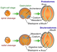
2) The reason why we, humans, can not help but start projects ass-backwards is because we are deuterostomes: we are simply made that way (see below).


Fig. 1. Deuterostomes vs protostomes. Credit: wikipedia

Wednesday, February 16, 2011

Abort! Abort!

ResearchBlogging.org


Sometimes things go so wrong that it is just easier to start all over again. Bacteria have these situations too - it's not just us, humans! - and the central dogma of molecular biology (DNA replication, transcription and translation) is no exception.

In essence all the three steps of the central dogma share the very same basic topology: there is a message that gets read, there is a tool that reads it and there is a product. It looks like so:

Say, in the case of translation mRNA (the message) gets read by the ribosome (the tool) and protein (the product) is produced. And when things go wrong, there are three things you can abort: the message, the product and the tool. Let us see how it goes.


Replication

DNA polymerase (the tool) reads the DNA (the message) and produces DNA (the product). And when wrong nucleotide is incorporated, DNA polymerase can excise it and continue making the product using so called  proof-reading mechanism. Complete abortion of the growing DNA strand does not happen, and if mistake is done, it is done and you live with it. Surely, there are ways to fix it later (recombination and so on), but not on the spot, during the replication.

Transcription


RNA polymerases can proof-read too. However, many more things can be done. Special set of transcription factors, called GreA and GreB in bacteria and TFSII in eucaryotes, can activate intrinsic hydrolytic activity of the RNA polymerase and cleave off the growing product. Stalled complex is resolved and now we can try again.

Translation


First, there is a proof-reading mechanism, but rather than cutting off the mis-incorporated letter, GTP is hydrolyzed by GTPase EF-Tu which brings the aminoacyl-tRNA.

Second, if the mistake is done, and wrong amino acid was incorporated after all, bacterial class-1 release factors RF1 and RF2 become prone to peptide-release independent of the stop codon, thus removing the product (the growing protein chain). In mitochondria translational system is bacterial-like, but much more insane, and several (as many as 4 in humans!) class-1 release factors are present, with some of them lacking the ability to recognize the stop codon at all (ICT1, for example), and these resolve stalled ribosomal complexes by cutting off the peptide as well as their bacterial counterparts.

Third, bacterial toxins such RelE and the like are resolving ribosomal complexes by cutting the message (mRNA) rather than the product. Calling them toxins is rather misguiding, they are more of the rescue factors.

And lastly, eukaryotic translational factors Dom34 and Hbs1 (related to termination factors eRF1 and eRF3) are splitting the stalled ribosome into subunits, re-setting the tool.

So it seems the further we move from the DNA, the more dispensable the production complex becomes: in the case of DNA polymerases we have only proof-reading, RNA polymerases can do that and also cleave the message, and translational machinery can do it all: cutting the message (RelE), cutting the product (release factors) and resetting the tool by splitting the ribosome into subunits (Dom34 and Hbs).

References:

Borukhov S, Sagitov V, & Goldfarb A (1993). Transcript cleavage factors from E. coli. Cell, 72 (3), 459-66 PMID: 8431948

Toulmé F, Mosrin-Huaman C, Sparkowski J, Das A, Leng M, & Rahmouni AR (2000). GreA and GreB proteins revive backtracked RNA polymerase in vivo by promoting transcript trimming. The EMBO journal, 19 (24), 6853-9 PMID: 11118220

Pedersen K, Zavialov AV, Pavlov MY, Elf J, Gerdes K, & Ehrenberg M (2003). The bacterial toxin RelE displays codon-specific cleavage of mRNAs in the ribosomal A site. Cell, 112 (1), 131-40 PMID: 12526800

Orlova M, Newlands J, Das A, Goldfarb A, & Borukhov S (1995). Intrinsic transcript cleavage activity of RNA polymerase. Proceedings of the National Academy of Sciences of the United States of America, 92 (10), 4596-600 PMID: 7538676

Kassavetis GA, & Geiduschek EP (1993). RNA polymerase marching backward. Science (New York, N.Y.), 259 (5097), 944-5 PMID: 7679800

Richter R, Rorbach J, Pajak A, Smith PM, Wessels HJ, Huynen MA, Smeitink JA, Lightowlers RN, & Chrzanowska-Lightowlers ZM (2010). A functional peptidyl-tRNA hydrolase, ICT1, has been recruited into the human mitochondrial ribosome. The EMBO journal, 29 (6), 1116-25 PMID: 20186120

Shoemaker CJ, Eyler DE, & Green R (2010). Dom34:Hbs1 promotes subunit dissociation and peptidyl-tRNA drop-off to initiate no-go decay. Science (New York, N.Y.), 330 (6002), 369-72 PMID: 20947765

Atkinson GC, Baldauf SL, & Hauryliuk V (2008). Evolution of nonstop, no-go and nonsense-mediated mRNA decay and their termination factor-derived components. BMC evolutionary biology, 8 PMID: 18947425

Antonicka H, Ostergaard E, Sasarman F, Weraarpachai W, Wibrand F, Pedersen AM, Rodenburg RJ, van der Knaap MS, Smeitink JA, Chrzanowska-Lightowlers ZM, & Shoubridge EA (2010). Mutations in C12orf65 in patients with encephalomyopathy and a mitochondrial translation defect. American journal of human genetics, 87 (1), 115-22 PMID: 20598281

Zaher HS, & Green R (2009). Quality control by the ribosome following peptide bond formation. Nature, 457 (7226), 161-6 PMID: 19092806