Wednesday, March 14, 2012

Proline residue in L11 as a key regulator of translational GTPases?

The ribosome is run by translational GTPases. Translational GTPases, in their turn, are regulated by the ribosome. They all bind in the same region (GAC, GTPase associated center) of the ribosome. In bacteria the GTPase binding site consists of a couple of rRNA elements: SRL (sarcin-ricin loop) and thiostrepton loop and several ribosomal proteins:L7/L12 stalk (L10 and L7/L12) and L11.

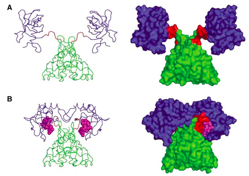
The latter is the main hero of a fresh paper in Nature Structural and Molecular Biology by Wang and coworkers. They show that bacterial translational GTPases (such as EF-G) when binding to the ribosome act as peptidyl-prolyl cis-trans isomerases (PPIases) driving isomerisation in the conserved residue in the ribosomal protein L11. This isomerisation, in turn, transmits signal to the ribosomal protein L7 /L12 - something that is necessary for efficient GTPase cycling on the ribosome.

I like L11 - it is a key protein for stringent response, and without it stringent factor RelA does not work, as was shown using E. coli mutants lacking L11 (Dabbs J. Bac 1979). These mutants are perfectly viable, but grow ten times slower then the wild type E. coli, most probably due to defects in the ribosome assembly (Hampl et al. JBC 1981). The very viability of the L11 knock-out strains tells us that L11 is not the key for keeping the ribosome running. In fact, less than a half of ribosomal proteins can be knocked-out in E. coli (22 out of 54, Shoji et al. JMB 2011), making L11 one of the less-important ones... and keeping an eye of the translational GTPases is definitely not one of the less-important functions!

This seems to be bit paradoxical - a ribosomal protein that is dispensable involved in something that is very central for protein biosynthesis. It gets even more fascinating when you look at the evolutionary aspect of the story (Gem Atkinson does that in her blog). Wang and colleagues managed to map  the PPIase site of EF-G.  As they show PPIase activity is universal for all the bacterial translational GTPases they tested, and the PPIase site is, surprisingly, quite a variable region of the G domain! So, do they all reinvent the weel separately? This is all most peculiar.

References:

Wang et al. A conserved proline switch on the ribosome facilitates the recruitment and binding of trGTPases. Nat Struct Mol Biol (2012) PIMD: 22407015

Tuesday, March 13, 2012

ppGpp induces production of fruiting bodies in Myxococcus xanthus

This post was chosen as an Editor's Selection for ResearchBlogging.org
E. coli is boring, admit it. At least in comparison with Myxococcus xanthus: a self-organized, predatory saprotrophic single-species biofilm called a swarm according to the Wikipedia. Now that sounds exciting! I wish one day somebody would call me "a self-organized, predatory biofilm called a swarm"! That would make a lovely email signature: "Vasili Hauryliuk, PhD,  self-organized, predatory biofilm called a swarm". Hell yes.

But I digress. Stringent response (or, to be more specific, RelA-mediated production of alarmone molecule ppGpp) regulates loads of things in bacterial physiology: it turns on bacterial survival mode and shuts down production of ribosomes, it induces virulence (cornered bacteria are deadly) and makes bugs more resistant to antibiotics. Now let us just imagine for a moment what stringent response can do to a "a self-organized, predatory biofilm called a swarm"! Exactly that was investigated in recent paper by Konovalova and colleagues (Konovalova et al. Mol Microbiology 2012).

Unlike boring E. coliMyxococcus xanthus has a life cycle (Fig. 1). It can swarm happily gobbling up other bacteria, or, if food supply is low, it can form a fruiting body (a life-stile similar to that of slime molds who are not bacteria but eucaryotes).


Fig. 1. 
Life cycle of a self-organized, predatory biofilm called a swarm (AKA Myxococcus xanthus).

Formation of the fruiting bodies depends on the functionality of the stringent response system (Harris et al. Gens Dev. 1998). How Konovalova and colleagues fill in the molecular details presenting an example of post-translational activation of secretion by regulated proteolysis.  Here is how it works.

Formation of the fruiting bodies depends on the cell-to-cell signaling, and this process, obviously, happens outside of the cell. It involves proteolysis of several extracellular target proteins by a subtilisin-like protease PopC, which needs to be exported outside of the cell in order to do its job. So now it turns out that RelA, working together with PopD protein, regulates PopC export, which is activated during starvation (and, therefore, production of ppGpp). The PopD:PolC complex formation is not affected by ppGpp, suggesting that regulation of export by RelA is using some indirect mechanism. And indeed, PopD turned out to be degraded during starvation in a FtsH-dependent manner, releasing PopC - a story somewhat similar to regulation of toxin:antitoxin pairs via antitoxin degradation by Lon protease during nutritional stress.

All this brings us to the question of importance of the regulated proteolysis during the stringent response. One known example of ppGpp-mediated control via protein degradation is degradation of ribosomal proteins by Lon protease induced by accumulation of polyphosphate. Unfortunately, usually stringent response on the whole-cell level is studied on the mRNA level, by, say, microarrays. It would be most educational to compare the changes on the mRNA level with changes on the proteome level and to pick up the protein degradation-mediated regulation pathways.

References:

Harris BZ, Kaiser D, & Singer M (1998). The guanosine nucleotide (p)ppGpp initiates development and A-factor production in Myxococcus xanthus. Genes & Development, 12 (7), 1022-35 PMID: 9531539

Konovalova A, Löbach S, & Søgaard-Andersen L (2012). A RelA-dependent two-tiered regulated proteolysis cascade controls synthesis of a contact-dependent intercellular signal in Myxococcus xanthus.  Molecular Microbiology  PMID: 22404381

Double life of mitochondrial ribosomal protein L7 12

Mitochondria have their own transcriptional and translational apparatus, even though they produce only a handful of proteins, therefore most of the proteins are imported from the cytoplasm. Trancription, translation and protein insertion into the membrane are interconnected: translational activators regulating mitochondrial translation are interacting with mitochondrial RNA polymerase via Nam1p and Sls1p proteins (Bryan et al. Genetics 2002), Puf proteins connect cytoplasmic translation and protein import into mitochondria by direct interaction with Tom20 subunit of the TOM protein import channel (Saint-Georges et al. PLoS ONE 2008).

But this seems not tight enough interaction for mitochondrial translation and transcription. It turnes out what mitohondrial ribosomal protein L7 12 (the one that brings translational GTPases to the ribosome), has a double life. Apart from doing its normal job as a part of the ribosome, it doubles as a transctiptional factor, selectively associating with human mitochondrial RNA polymerase and activating it (Surovtseva et al. PNAS 2011). And as if it is not enough, there are several paralogues of L7 12 in mitochondria, both in plants (Delage et al. Biochimie 2007) and in mammals (Koc et al. JBC 2001). 

Monday, March 12, 2012

Measuring nucleotide concentrations inside the living cells

ResearchBlogging.org
Taking biological system apart and doing experiments in vitro is a very powerful approach. However, Nature has loads of dirty tricks up her sleeve, so doing experiments in vivo is more kosher - at least you get all the concentrations rights and will have all of the components present in the system.

Cells use a whole plethora of nucleotide-based messengers (Pesavento and Hengge, Curr. Opin. Microbiol. 2009), and following concentrations of these in vivo is something microbiologists would love to do. It is possible for some, and  c-di-GMP is an example. This nucleotide binds to numerous targets, and one of them is PilZ proteins. When binding to PilZ domain, c-di-GMP promotes a massive structural rearrangement, and this interaction can be monitored by adding a FRET pair to PilZ (Benach et al. EMBO J 2007) (Fig. 1). FRET response can be converted in c-di-GMP concentration using a calibration curve, and - viola! - c-di-GMP can be measured in the individual live cells in real time using a PilZ-GFP-based FRET detector (Christen et al. Science 2010).


Fig. 1. PlzD: Apo (A) and in complex with c-di-GMP (figure from Benach et al. EMBO J 2007).

The problem with this approach is that is far from being universal. First, one has to have a protein that binds your target nucleotide and undergoes massive rearrangements. Second, this protein should be nice enough to work with so that you can add two GFP molecules to it to make a FRET pair, and still be able to express the protein for in vitro work (one needs to calibrate the FRET response, right?). In the case of stringent response there seem to be no such proteins for detection of my favorite nucleotide, ppGpp... too bad!

Well, there seems to be a new method out there, and this one holds great promise. In their recent Science paper Paige and colleagues use an RNA-based FRET pair using RNA mimic of GFP (Paige et al. Science 2011) combined with a small-molecule-specific aptamer (Fig. 2). When the ligand binds, RNA forms a stable structure and FRET is on! They have followed in E. coli concentrations of two molecules - ADP and SAM. However, aptamers can be evolved for other targets, and this makes this method potentially applicable for detecting whatever molecule that picks your fancy.


Fig. 2. Schematic representartion of the aptamer-based FRET sensor for in vivo detection of small molecules (figure from Paige et al. Science 2012).

References:

Paige JS, Nguyen-Duc T, Song W, & Jaffrey SR (2012). Fluorescence imaging of cellular metabolites with RNA. Science (New York, N.Y.), 335 (6073) PMID: 22403384

Paige JS, Wu KY, & Jaffrey SR (2011). RNA mimics of green fluorescent protein. Science (New York, N.Y.), 333 (6042), 642-6 PMID: 21798953 Christen M, Kulasekara HD, Christen B, Kulasekara BR, Hoffman LR, & Miller SI (2010). 


Matthias Christen, Hemantha D Kulasekara, Beat Christen, Bridget R Kulasekara, Lucas R Hoffman, and Samuel I Miller (2010) Asymmetrical distribution of the second messenger c-di-GMP upon bacterial cell division. Science (New York, N.Y.), 328 (5983), 1295-7 PMID: 20522779

 Benach J, Swaminathan SS, Tamayo R, Handelman SK, Folta-Stogniew E, Ramos JE, Forouhar F, Neely H, Seetharaman J, Camilli A, & Hunt JF (2007). The structural basis of cyclic diguanylate signal transduction by PilZ domains. The EMBO journal, 26 (24), 5153-66 PMID: 18034161